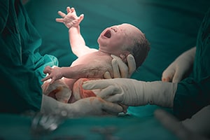
A congenital defect or Birth defect is a visible or internal or chemical abnormality of the children caused by genetics, infection, radiation, or drug exposure, or of unknown reason. Examples include sickle cell anemia, phenyl ketonuria, and Down syndrome. The defect can affect any part of the child’s body, which can be:
- Clearly Visible like a missing arm or a birthmark.
- Internal (inside the body), like a defective kidney or a ventricular septal defect (a hole between the lower chambers of the baby’s heart).
- A chemical imbalance, like phenylketonuria (a defect in a chemical reaction that results in developmental delay).
The baby can be born with one birth defect such as a cleft lip (a gap in their upper lip) or multiple birth defects such as a cleft lip and cleft palate (a hole in the roof of their mouth) together, or even a cleft lip and cleft palate with defects of the brain, heart and kidneys.
The Doctor may not be able to detect all birth defects right when the baby is born. Some defects, such as scoliosis, might not be apparent until the child is several months old. An abnormal kidney might take years to be discovered. 2-3% of infants have one or more defects at birth. That number increases to 5% by age one.
OTHER EXAMPLES OF BIRTH DEFECTS

- Duodenal atresia, an obstruction in the small intestine. It can cause polyhydramios (extra fluid around the baby in pregnancy), which can increase the risk of preterm birth. It is sometimes associated with other genetic syndromes.
- Dandy walker malformation, an abnormal development of the posterior fossa (a space in baby’s skull) and cerebellum (a section of the brain). This can cause a variety of problems.
- Limb defects, which happen when the fetal amnion (the inner lining of the amniotic sack) wraps around parts of the fetus (like a finger or foot).
CAUSES
The reasons of the defects may be:
- Genetic or hereditary factors.
- Environmental factors like infections during pregnancy.
- Exposure to some drugs during pregnancy.
1.HEREDITARY FACTORS
About 20% of birth defects are caused by genetic or hereditary factors. Genetic causes fall into three general categories:
- Chromosomal abnormalities.
- Single-gene defects.
- Multifactorial.
Every human body cell contains 46 chromosomes, and each chromosome contains thousands of genes. Each gene contains a blue print that controls the functions or development of a specific part of the body. People who have either too many or too few chromosomes will therefore receive a scrambled message regarding anatomic development and function.
Down syndrome is an example of a condition caused by too many chromosomes. Because of an accident during cell division, individuals with Down syndrome have an extra copy of a particular chromosome (chromosome 21). This extra chromosome can cause a typical constellation of birth defects.
Characteristic features of Down syndrome include developmental delay, muscle weakness, downward slant of the eyes, low-set and malformed ears, an abnormal crease in the palm of the hand and birth defects of the heart and intestines.
With Turner syndrome, a female lacks part or all of one X chromosome. In the affected women, this can cause short stature, learning disabilities and absence of ovaries.
Trisomy 13 (Patau Syndrome) and Trisomy 18 (Edwards Syndrome) are caused by inheriting extra copies of the 13th or 18th chromosome, respectively. These are rarer, more serious conditions which cause severe birth defects that are incompatible with survival after birth.
In addition to inheriting an extra or absent chromosome, deletions or duplications of single genes can also cause developmental disorders and birth defects. One example is cystic fibrosis (a disorder that causes progressive damage of the lungs and pancreas).
Defective genes can also be caused by accidental damage, a condition called “sponta neous mutation.” Most cases of achondroplasia (a condition that causes extreme short stature and malformed bones) are caused by new damage to the controlling gene. In addition, recombination errors can cause translocations of chromosomes which may lead to complex birth defects.
2,ENVIRONMENTAL FACTORS LIKE INFECTION
Environmental factors can increase the risk of miscarriage, birth defects, or they might have no effect on the baby at all, depending on at what point during the pregnancy the exposure occurs. The baby goes through two major stages of development after conception. The first, or embryo stage, occurs during the first 10 weeks after conception. Most of the major body systems and organs form during this time.
The second, or fetal stage, is the remainder of the pregnancy. This fetal period is a time of growth of the organs and of the fetus in general. The developing baby is most vulnerable to injury during the embryo stage when organs are developing. Infections and drugs can cause the greatest damage when exposure occurs two to 10 weeks after conception. Diabetes and obesity can increase the child’s risk of birth defects.
3.EXPOSURE TO DRUGS
Some medicines and recreational drugs can cause birth defects, which are most severe when used during the first three months of pregnancy. Thalidomide, an anti-nausea medicine prescribed during the 1960s, caused birth defects called phocomelia (absence of most of the arm with the hands extending flipper-like from the shoulders).
Sotretinoin or Roaccutane which is a retinoid, a synthetic form of vitamin A used to treat skin conditions can cause fetal retinoid syndrome. Its Characteristics include delayed growth, malformations of the skull and face, abnormalities of the Central Nervous System. Heart, Parathyroid gland, the Renal & Thymus gland.
Consumption of drugs containing alcohol can also causes birth defects. The typical defect caused by the mother’s alcohol use can lead to Fetal alcohol syndrome with learning disabilities. developmental delay, irritability, hyperactivity. poor coordination, abnormalities of facial features etc.
Another environmental factor is uterine constraint. The fetus grows in the mother’s uterus and is surrounded by amniotic fluid (similar to being suspended in a bag of water) that cushions it from excessive pressure. If the sack of fibers that holds the fluid breaks, bands of fibers from the torn sack can press on the fetus and cause amniotic band syndrome (which can result in partial contraction of or amputation of an arm or leg). An inadequate amount of amniotic fluid can cause excessive pressure on the entire baby, causing pulmonary hypoplasia (lack of development of the lungs).
Only 30% of birth defects are identified by the Medical science and the rest remain without a straightforward cause. As many as 50-70% of birth defects are sporadic, and their cause remains unknown. A combination of environmental and genetic factors can increase the risk of certain birth defects.
Stress is also a leading cause, results in increased catecholamine production, which in turn leads to decreased uterine blood flow and increased fetal hypoxia which may affects a variety of developmental processes.
DIAGNOSIS AND TESTS

These defects can be diagnosed before the child’s birth, at birth and after birth. Most are found within child’s first year. During pregnancy screening with ultrasounds or blood tests can identify birth defects and genetic disorders. If the screening test shows some abnormality, a diagnostic test is often recommended.
First trimester screenings check for problems with baby’s heart, and for chromosomal disorders such as Down syndrome. The tests include:
- Maternal blood tests can measure protein levels or circulating free fetal DNA in maternal blood. Abnormal results can indicate increased risk of a fetal chromosomal disorder.
- An ultrasound looks for extra fluid behind the baby’s neck. That is indicative of risk of a heart defect or chromosomal disorder. Second trimester screenings check for problems with the structure of the baby’s anatomy. The tests include:
- • Maternal serum tests in the second trimester can screen for chromosomal disorders and/or spina bifida.
- An anomaly ultrasound checks the size of the baby and checks for birth defects.
- • More tests may be recommended if a screening test is abnormal. Such diagnostic tests are also offered to women with higher risk pregnancies. The tests include:
- Fetal echocardiogram This ultrasound is focused on the baby’s heart, and may be done in certain high risk pregnancies, or if a heart defect is suspected on anomaly ultrasound. Not all heart defects can be seen before the birth.
- Fetal MRI is ordered in cases of suspected birth defects of the baby’s brain or nervous system.
- Chorionic villus sampling Here a very small piece of the placenta is collected and will be examined for chromosomal or genetic disorders.
- Amniocentesis A small amount of amniotic fluid is collected and the cells are tested for chromosomal disorders and genetic problems like cystic fibrosis or Tay-Sachs disease. Amniocentesis can also test for certain infections such as cytomegalovirus (CMV).
Many birth defects may not be diagnosed until after the baby is born. They may be seen immediately, like a cleft lip, or diagnosed later.
TREATMENT AND PREVENTION

Treatment is done based on the diagnosis. Most birth defects cannot be prevented. But can take some important steps to promote a healthy pregnancy. These include:
- Frequent medical check up
- Take a prenatal vitamin with 400 mcg of folic acid, if trying to conceive, or are sexually active and not using contraception.
- Contact the Gynecologist immediately after confirmation of pregnancy.
- Keep diabetes under control. Poor control of diabetes during pregnancy increases the chances for birth defects and other problems for the pregnancy.
- Maintain a healthy weight with body mass index below 30 to avoid complications during pregnancy.
- Abstain from usage of alcoholic drinks, psychotropic and narcotic drugs and smoking.
- Inform the doctor regarding the medicines and supplements taken during pregnancy.
BEST FOOD DURING PREGNANCY

- Lean meat, poultry, fish, and eggs which are good sources of protein.
- Other options include beans, tofu, dairy products, and peanut butter.
- Breads and grains: Mothers should choose grains that are high in fiber and enriched such as whole-grain breads, cereals, pasta, and rice


